Figure 3.

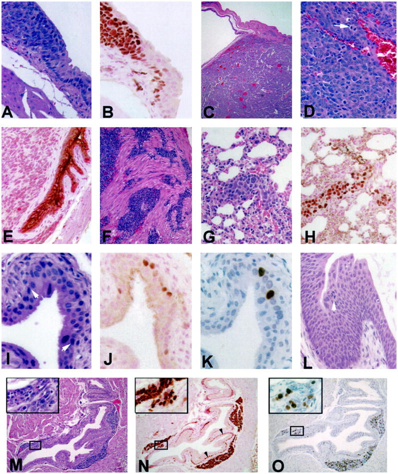
Bladder lesions and their progression in CK19-TAg transgenic mice. A: Transition from normal-appearing urothelium to neoplastic urothelium in a 12-week-old CK19-TAg transgenic mouse. B: Tissue section adjacent to A displaying immunohistochemical localization of TAg protein. C: Bladder tumor from a 12-week-old CK19-TAg transgenic mouse. D: Higher power photomicrograph of a bladder neoplasm illustrating typical cellular characteristics, including the presence of mitotic figures (arrow). E: Transgenic mouse urothelium displaying localization of uroplakin protein, which is excluded from neoplastic cells. F: Invasion of neoplastic urothelial cells into muscle. G: H&E stain of lung metastasis in the vessel of a 12-week-old CK19-TAg transgenic mouse, which expresses TAg protein (H). I−K: Earliest urothelial lesion visualized in a 3-week-old mouse. In I, arrowheads indicate the approximate lesion margins. There is an increase of nuclear TAg protein in some cells in the lesion (J), and an associated increase in BrdU labeling (darkly labeled cells in K). L: Large CIS present in a 6-week-old transgenic mouse. Arrowhead indicates a tripolar mitosis. M−O: H&E stain (M), TAg protein localization (N), and BrdU localization (O) in bladder of a 9-week-old transgenic mouse displaying characteristic nests of neoplastic cells between the suburothelial stroma and muscle. Insets display magnified views of the boxed areas in which a focal collection of cells extends through the stroma, appearing to connect urothelium and the substromal neoplastic focus of cells. Arrowheads in N indicate TAg-expressing hypertrophied basal urothelial cells in this mouse. For TAg and uroplakin immunohistochemistry, counterstain is nuclear-fast red; for BrdU immunohistochemistry, counterstain is hematoxylin. All others are H&E. Original magnifications, ×400 (A, B, D, E, G−L, and insets for M−O); ×40 (C); and ×100 (F, M−O).
