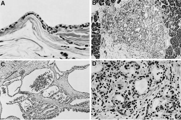
Figure 1.

Histology of multiple pancreatic cystic lesions in VHL patients (H&E). A: Benign serous cyst (×400). B: Microscopic MCA (×200). C: Macroscopic MCA (×200). D: High power view of microscopic MCA (×400); epithelial cells intermixed with numerous endothelial cells that form small vessels, and stromal fibrosis.
