Figure 3.

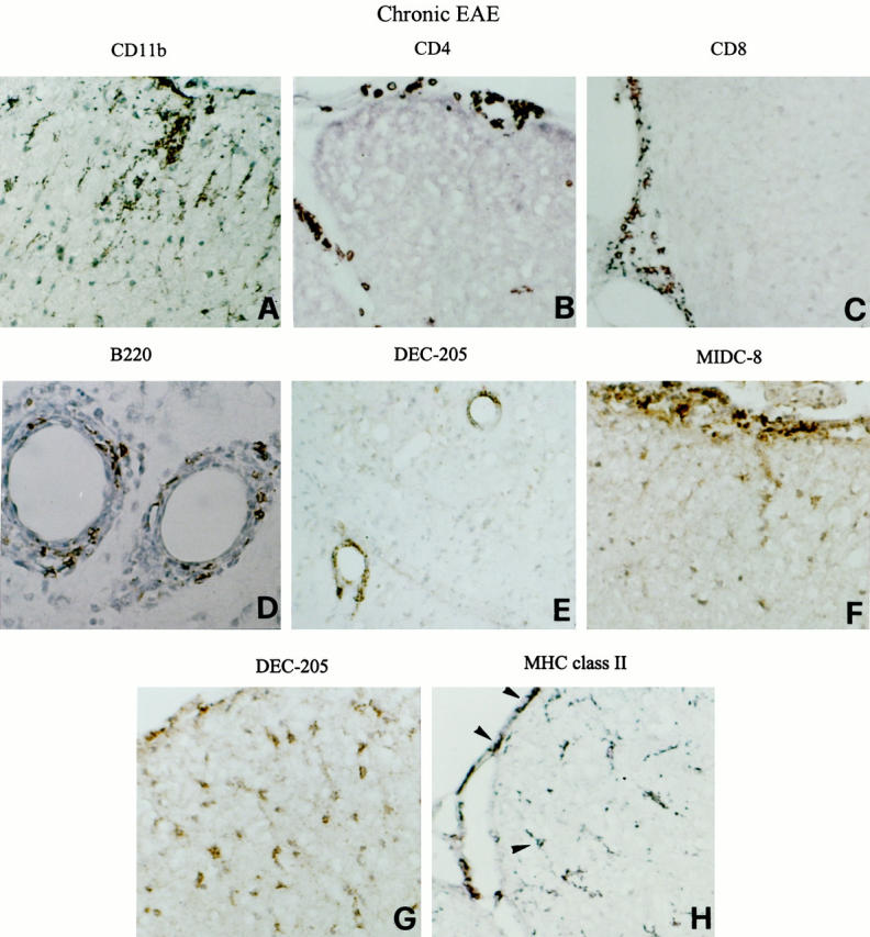
Persistence of mature DCs in the CNS of mice with chronic EAE. Representative thoracic (A, B, F, G, and H) and cervical (C) spinal cord and cerebellar (D and E) sections from a SJL mouse with chronic EAE (grade 3) are shown. CNS sections were stained for the indicated markers and visualized with DAB, as described in Materials and Methods. The immune infiltrates remain primarily confined to the meninges and around blood vessels and are composed of CD11b+ macrophages (A), CD4+ T cells (B), CD8+ T cells (C), as well as B220+ B cells (D). Cells positive for DEC-205 (E) and for the mature DC marker MIDC-8 (F) accumulate perivascularly and in the meninges. Anti-DEC-205 mAb stains process-bearing cells within the dorsal spinal cord white matter (G). Some of the MHC class II+ cells detected in the spinal cord meninges and white matter (H) have a DC-like morphology (arrowheads). Original magnifications: ×500 (A–C and E–H), and ×1000 (D).
