
Figure 8.
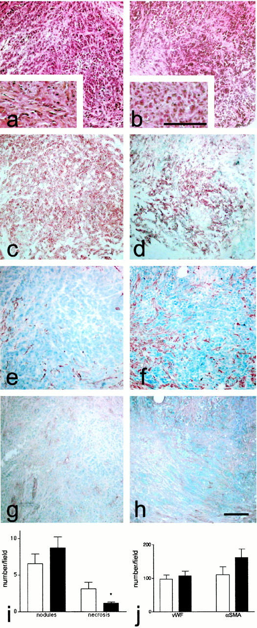
Morphology of the colon tumors in the experimental rat model. Immunohistochemistry was performed in consecutive sections of tumors from control (a, c, e, and g) and bosentan-treated rats (b, d, f, and h). Photographs are presented in bright-field illumination. Eosin-hematoxylin-safran staining (a and b) was performed for assessing the tumor appearance. Collagen characterized with the safran staining (orange) was shown at higher magnification (insert in a and b). Tumor epithelial cells bound anti-keratin antibody (c and d). Smooth muscle cells were identified with anti-α-SMA (e and f) and endothelial cells with an anti-vWF antibody (g and h). Histological scores (i) were obtained by counting the number of necrotic areas and nodules in the field of the ×4 objective in control (white bars) and bosentan-treated animals (black bars). j: Distribution of specific markers in rat nodules. The cells stained for vWF and α-SMA were counted in the field of the ×40 objective in control (open bars) and bosentan-treated animals (black bars). The results averaged values of seven different samples that are representative of all of the specimens analyzed. Significant differences (P ≤ 0.05) are indicated by an asterisk. Scale bar, 100 μm.