
Figure 1.
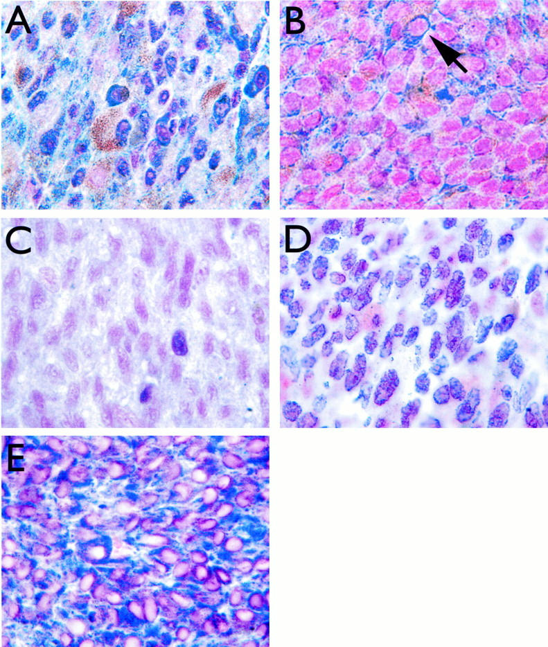
Immunohistochemical staining for Rb, p53, MDM2, and Bcl2. A: Strong nuclear staining for Rb in a uveal melanoma with no local treatment before enucleation. B: Staining for Rb in a uveal melanoma that failed brachytherapy before enucleation (patient no. 32). Note absence of nuclear staining and strong cytoplasmic staining (arrow). C: Weak staining for p53 in a representative uveal melanoma. D: Staining for MDM2 in a representative uveal melanoma, demonstrating strong nuclear expression in most cells. E: Staining for Bcl2 in a representative uveal melanoma, demonstrating strong cytoplasmic expression in most cells. Streptavidin-biotin-peroxidase method, Vector SG peroxidase substrate, and nuclear fast red counterstain (see Materials and Methods). Original magnification, ×100.