
Fig. 3.
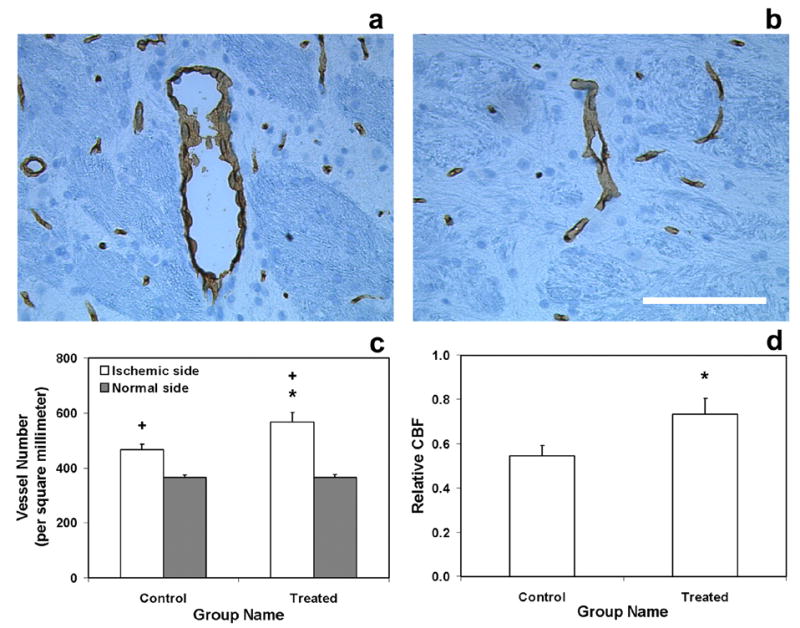
Immunoreactive cerebral vessels from a treated animal (a, ipsilateral side; b, contralateral side. Bar = 100μm), vessel density (c) and relative CBF (d) in lesion boundary area 6-weeks after stroke. Treatment with sildenafil significantly increased cerebral vessel number in the lesion boundary area compared with the control group (c). Relative CBF in the lesion boundary area was also significantly improved (d) compared with the control group. Significance of difference: * = p < 0.05, comparing treated and control groups in the ischemic boundary region; + = p < 0.05, comparing ischemic boundary region with the homologous area in the contralateral (normal) side of rat brain in treated group and control group, respectively.