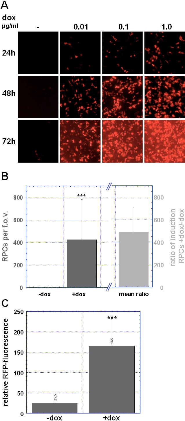
Figure 2. Doxycycline-induced RFP-expression.

(A) RFP-expression in culture mediated by HET6C-tk39. Fluorescence microscopy of RFP-expressing cells that were infected with HET6C-tk39 in the presence or absence of doxycycline. Although expression of RFP in HET6C-tk39 infected but untreated cells is very tightly regulated, we find some leakiness of the construct that is indicated by some single RFP-positive cells slightly visible at 72 h past infection. (B) Counting of RFP-positive cells (RPC) was performed+/−doxycycline (48 h post induction, 1 µg/ml). Columns in dark grey represent the number of RPCs/f.o.v., column in light grey represents the mean ratio of induction (number of RFP-positive cells in presence or absence of doxycycline). Histograms represent the calculated means+/−SD. (C) Relative intensity of red fluorescence was recorded in single cells by means of a ROI analysis using MPI-Tool imaging software, error bars signify the SD. ***, P<0.001 compared to non-induced cells (Mann-Whitney Rank Sum Test).
