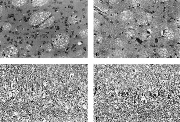
Figure 2.
Light microphotographs demonstrating the effect of the NMDA antagonist CPP on slowly progressing neurodegeneration induced in the striatum by systemic administration of 3NP and in the hippocampus by traumatic brain injury. (A) Morphology of striatum after treatment with 3NP, 12 mg/kg per d over 28 days, 3 days after termination of treatment. No obvious neurodegeneration can be detected in the striatum; the spiny neurons have normal appearance. (B) Profound neuronal loss and predominance of glia in the striatum 3 days after termination of treatment with 3NP, 12 mg/kg per d, and CPP, 24 mg/kg per d over 28 days. Large-size striatal neurons are relatively preserved. (C) Hippocampal pathology in the CA3 subfield 3 days after traumatic brain injury. Dark argyrophylic profiles indicate ongoing degeneration in pyramidal layer. Intact pyramidal cells with prominent nuclei are also present. (D) The effect of treatment with CPP, 3 × 30 mg/kg given i.p. 1, 2, and 3 h after trauma is shown. Widespread degeneration of pyramidal neurons predominates. Magnifications: A and B, ×60 (cresyl violet stain); C and D, ×80 (Fink and Heimer stain).

