
Figure 3.
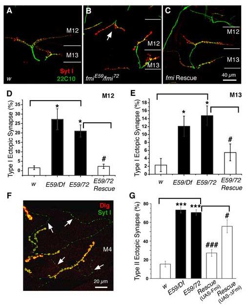
Mutations in fmi cause a significant increase in the number of ectopic type I boutons
(A-C). Representative immunocytochemical staining of the NMJs on muscles 12 and 13 with antibodies to the synaptic vesicle protein synaptotagmin I (Syt I, red) and the microtubule-associated protein Futsch (22C10, green) in the control (w, A), the fmiE59/ fmi72 (B), and rescued fmiE59/ fmi72 (C) larvae. Ectopic type I synapses on muscle 12 are indicated by the arrows in panel (B). The while lines mark the boundary between muscles 12 and 13.
(D & E). Histograms show that fmi mutations significantly increase the fraction of muscles (muscle 12, D and muscle 13, E) receiving ectopic type I synapses compared to that in the w control larvae (* p<0.05). This defect is effectively rescued by neuronal expression of the wild type Flamingo in the fmiE59/ fmi72 mutant background (# p<0.05).
(F). A representative image showing ectopic type II innervations on muscle 4 in fmi mutants labeled with the postsynaptic marker Dlg (red) and presynaptic marker Syt I (green). Type II synaptic boutons are marked by arrows.
(G). Histogram plots show that the percentage of muscle 4 receiving ectopic type II synaptic input is significantly increased in fmiE59/Df and fmiE59/ fmi72 mutant larvae (***P<0.001). This defect can be effectively rescued to near wild type levels by neuronal expression of the wild type Flamingo in the fmiE59/ fmi72 mutant background, but only partially rescued by expression of the truncated Flamingo (### p<0.001; # P<0.05).