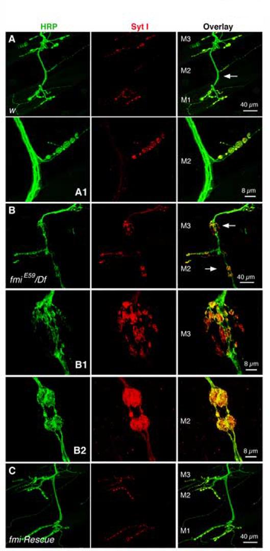
Figure 4.

En passant synapses are found on muscles 2 and 3 in the fmi mutant
(A & A1). Representative images of axons (HRP, green) and synapses (Syt I, red) on muscles 1, 2 and 3 in the control larvae (A). The NMJ indicated by the arrow is shown at higher magnification in panel (A1). Note the usual ‘beads-on-a-string’ pattern of synaptic boutons extended on the muscle surface.
(B-B2). Representative images of axons (HRP, green) and synapses (Syt I, red) on muscles 2 and 3 in the fmiE59/Df larvae (B). The NMJs indicated by the arrows are shown at higher magnification below in panels (B1) and (B2). Compared to the NMJ in the control larva, these axons arrive on muscles 2 and 3 as one bundle, then defasciculate into individual axons and form boutons along the axon on muscles 2 and 3, and finally converge back to one nerve bundle and proceed to the next muscle. Synaptic vesicle proteins represented by Syt I are well retained within each en passant synapse. Note the irregular sized, often enlarged, synaptic boutons.
(C). The formation of these en passant synapses can be partially suppressed by neuronal expression of the wild type Flamingo in the mutant background.
