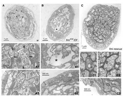
Figure 8.

Axonal degeneration of segmental nerves in the fmi mutant larvae and its rescue by neuronal expression of Flamingo
(A-A2). Panel (A) shows a representative cross-section of the segmental nerve from a control (w) larva. The arrowheads point to one axon that is further magnified in panel (A1). Axons are packed with mitochondria and microtubules. The edge of the segmental nerve is magnified in panel (A2) to illustrate the thickness of the neuronal sheath (double headed arrows).
(B-B2). Panel (B) shows a representative cross-section of the segmental nerve for an fmiE59/Df mutant larva. In contrast to panel (A), the mutant nerve has more empty ‘holes’ and is relatively less packed with axons and glial processes. The asterisk marks the gap in the nerve that is magnified in panel (B1). These axons are less packed with mitochondria and microtubules. Double-headed arrows in panel (B2) delimit the sheath surrounding the segmental nerves of the mutant. Note the thickening of the nerve sheath.
(C-C3). Panel (C) shows a representative cross-section of the segmental nerve for an fmiE59/Df mutant larva rescued by expressing a wild type Flamingo in postmitotic neurons. In contrast to panel (B), the rescued nerve is larger in diameter and well packed with axons and glial processes. The asterisk marks one axon undergoing degeneration that is magnified in panel (C1). The arrowheads point to one axon that is further magnified in panel (C2). Similar to axons in the wild type, the rescued axons are packed with mitochondria and microtubules. Double-headed arrows in panel (C3) delimit the sheath surrounding the segmental nerves of the rescued larva. Note that the nerve sheath is restored to the thickness found in the wild type.
