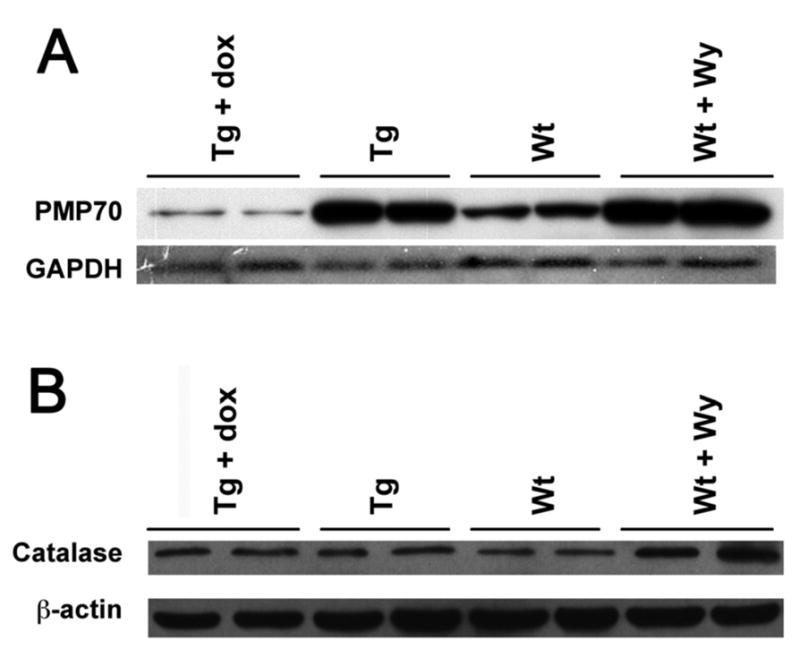
Figure 3. Induction of peroxisome proliferation in the LAP-VP16PPARα mice.

Western bolt analysis of peroxisome proliferation marker enzymes in liver total protein from 8–10 weeks old mice using antibody indicated. (A) PMP70 peroxsiomal membrane protein 70, (B) catalase. Note that both Wt mice treated with Wy-14,643 and LAP- VP16PPARα (Tg) mice in the absence of dox showed the increased of PMP70, however, only Wt mice treated with Wy-14,643 showed increased catalase protein expression.
