Abstract
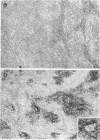
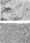
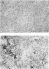
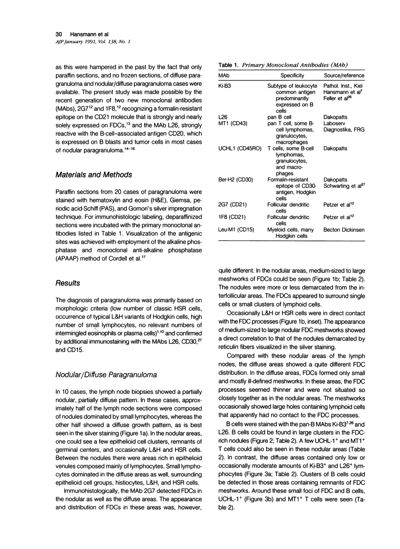
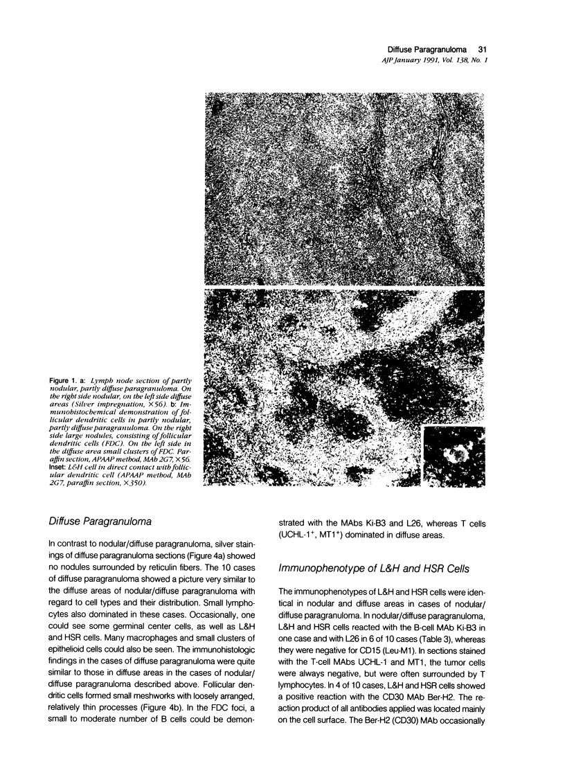
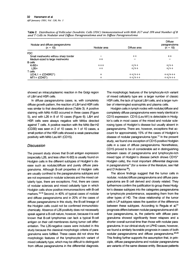
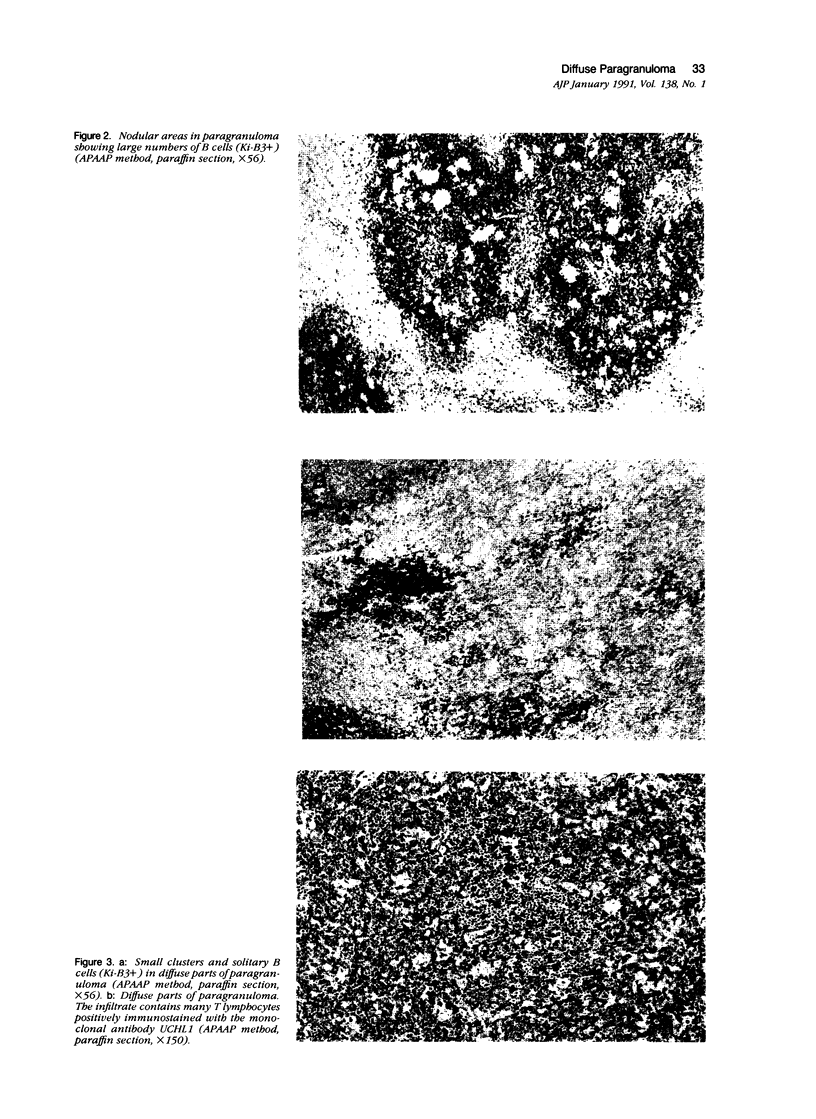
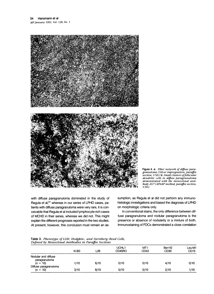
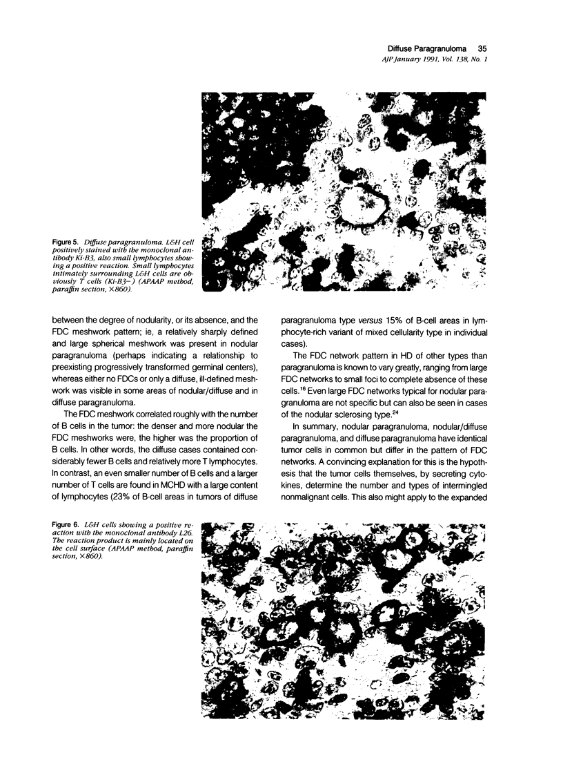
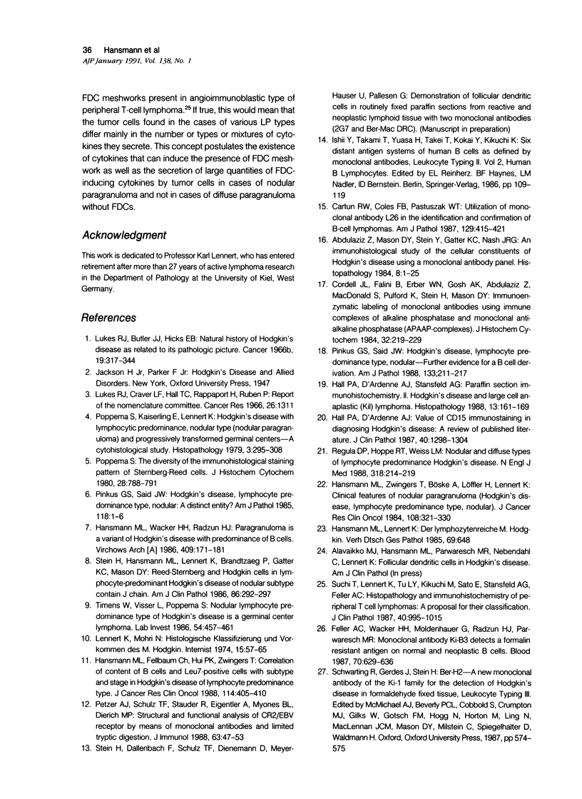
Lymph node sections from 10 cases of mixed nodular/diffuse and 10 cases of completely diffuse lymphocyte-predominant Hodgkin's disease (LPHD) were immunophenotyped. The results obtained were compared with those of nodular LPHD (nodular paragranuloma). In conventional stains, nodular/diffuse LPHD differed from diffuse LPHD in the presence of nodularity, which can be best demonstrated with silver impregnation. Immunohistologic analysis showed a correlation of the difference in nodularity with the presence or absence and pattern of follicular dendritic cell (FDC) meshwork, ie, a relatively sharply defined and large spherical meshwork was present in nodular areas of nodular/diffuse LPHD, whereas FDCs were either absent or present in a diffuse, ill-defined meshwork, usually of small size, in the diffuse zones of nodular/diffuse LPHD and in diffuse LPHD. The amount of FDC meshwork corresponded roughly to the number of reactive B cells and T cells, meaning that in diffuse areas significantly fewer B cells and more T cells were observed than in nodular areas. The immunohistologic analysis also showed that the antigen profile (positivity with the monoclonal B-cell marker L26 in the majority [14/20] of cases and negativity for CD15 in all but one of 20 cases) of the tumor cells in both nodular/diffuse LPHD and diffuse LPHD were comparable while it was different from the antigen profile (L26- and CD15+) in most cases of nodular sclerosis and mixed cellularity types of HD. This suggests that the considered subtypes of LPHD differ mainly in FDC pattern, but not in origin and nature of the tumor cells. This further justifies assignment of the above-mentioned LPHD subtypes to the category paragranuloma (LPHD).
Full text
PDF
Images in this article
Selected References
These references are in PubMed. This may not be the complete list of references from this article.
- Abdulaziz Z., Mason D. Y., Stein H., Gatter K. C., Nash J. R. An immunohistological study of the cellular constituents of Hodgkin's disease using a monoclonal antibody panel. Histopathology. 1984 Jan;8(1):1–25. doi: 10.1111/j.1365-2559.1984.tb02318.x. [DOI] [PubMed] [Google Scholar]
- Cartun R. W., Coles F. B., Pastuszak W. T. Utilization of monoclonal antibody L26 in the identification and confirmation of B-cell lymphomas. A sensitive and specific marker applicable to formalin-and B5-fixed, paraffin-embedded tissues. Am J Pathol. 1987 Dec;129(3):415–421. [PMC free article] [PubMed] [Google Scholar]
- Cordell J. L., Falini B., Erber W. N., Ghosh A. K., Abdulaziz Z., MacDonald S., Pulford K. A., Stein H., Mason D. Y. Immunoenzymatic labeling of monoclonal antibodies using immune complexes of alkaline phosphatase and monoclonal anti-alkaline phosphatase (APAAP complexes). J Histochem Cytochem. 1984 Feb;32(2):219–229. doi: 10.1177/32.2.6198355. [DOI] [PubMed] [Google Scholar]
- Feller A. C., Wacker H. H., Moldenhauer G., Radzun H. J., Parwaresch M. R. Monoclonal antibody Ki-B3 detects a formalin resistant antigen on normal and neoplastic B cells. Blood. 1987 Sep;70(3):629–636. [PubMed] [Google Scholar]
- Hall P. A., D'Ardenne A. J. Value of CD15 immunostaining in diagnosing Hodgkin's disease: a review of published literature. J Clin Pathol. 1987 Nov;40(11):1298–1304. doi: 10.1136/jcp.40.11.1298. [DOI] [PMC free article] [PubMed] [Google Scholar]
- Hall P. A., d'Ardenne A. J., Stansfeld A. G. Paraffin section immunohistochemistry. II. Hodgkin's disease and large cell anaplastic (Ki1) lymphoma. Histopathology. 1988 Aug;13(2):161–169. doi: 10.1111/j.1365-2559.1988.tb02021.x. [DOI] [PubMed] [Google Scholar]
- Hansmann M. L., Fellbaum C., Hui P. K., Zwingers T. Correlation of content of B cells and Leu7-positive cells with subtype and stage in lymphocyte predominance type Hodgkin's disease. J Cancer Res Clin Oncol. 1988;114(4):405–410. doi: 10.1007/BF02128186. [DOI] [PubMed] [Google Scholar]
- Hansmann M. L., Wacker H. H., Radzun H. J. Paragranuloma is a variant of Hodgkin's disease with predominance of B-cells. Virchows Arch A Pathol Anat Histopathol. 1986;409(2):171–181. doi: 10.1007/BF00708326. [DOI] [PubMed] [Google Scholar]
- Hansmann M. L., Zwingers T., Böske A., Löffler H., Lennert K. Clinical features of nodular paragranuloma (Hodgkin's disease, lymphocyte predominance type, nodular). J Cancer Res Clin Oncol. 1984;108(3):321–330. doi: 10.1007/BF00390466. [DOI] [PubMed] [Google Scholar]
- Lennert K., Mohri N. Histologische Klassifizierung und Vorkommen des M. Hodgkin. Internist (Berl) 1974 Feb;15(2):57–65. [PubMed] [Google Scholar]
- Petzer A. L., Schulz T. F., Stauder R., Eigentler A., Myones B. L., Dierich M. P. Structural and functional analysis of CR2/EBV receptor by means of monoclonal antibodies and limited tryptic digestion. Immunology. 1988 Jan;63(1):47–53. [PMC free article] [PubMed] [Google Scholar]
- Pinkus G. S., Said J. W. Hodgkin's disease, lymphocyte predominance type, nodular--a distinct entity? Unique staining profile for L&H variants of Reed-Sternberg cells defined by monoclonal antibodies to leukocyte common antigen, granulocyte-specific antigen, and B-cell-specific antigen. Am J Pathol. 1985 Jan;118(1):1–6. [PMC free article] [PubMed] [Google Scholar]
- Pinkus G. S., Said J. W. Hodgkin's disease, lymphocyte predominance type, nodular--further evidence for a B cell derivation. L & H variants of Reed-Sternberg cells express L26, a pan B cell marker. Am J Pathol. 1988 Nov;133(2):211–217. [PMC free article] [PubMed] [Google Scholar]
- Poppema S., Kaiserling E., Lennert K. Hodgkin's disease with lymphocytic predominance, nodular type (nodular paragranuloma) and progressively transformed germinal centres--a cytohistological study. Histopathology. 1979 Jul;3(4):295–308. doi: 10.1111/j.1365-2559.1979.tb03011.x. [DOI] [PubMed] [Google Scholar]
- Poppema S. The diversity of the immunohistological staining pattern of Sternberg-Reed cells. J Histochem Cytochem. 1980 Aug;28(8):788–791. doi: 10.1177/28.8.6777426. [DOI] [PubMed] [Google Scholar]
- Regula D. P., Jr, Hoppe R. T., Weiss L. M. Nodular and diffuse types of lymphocyte predominance Hodgkin's disease. N Engl J Med. 1988 Jan 28;318(4):214–219. doi: 10.1056/NEJM198801283180404. [DOI] [PubMed] [Google Scholar]
- Stein H., Hansmann M. L., Lennert K., Brandtzaeg P., Gatter K. C., Mason D. Y. Reed-Sternberg and Hodgkin cells in lymphocyte-predominant Hodgkin's disease of nodular subtype contain J chain. Am J Clin Pathol. 1986 Sep;86(3):292–297. doi: 10.1093/ajcp/86.3.292. [DOI] [PubMed] [Google Scholar]
- Suchi T., Lennert K., Tu L. Y., Kikuchi M., Sato E., Stansfeld A. G., Feller A. C. Histopathology and immunohistochemistry of peripheral T cell lymphomas: a proposal for their classification. J Clin Pathol. 1987 Sep;40(9):995–1015. doi: 10.1136/jcp.40.9.995. [DOI] [PMC free article] [PubMed] [Google Scholar]
- Timens W., Visser L., Poppema S. Nodular lymphocyte predominance type of Hodgkin's disease is a germinal center lymphoma. Lab Invest. 1986 Apr;54(4):457–461. [PubMed] [Google Scholar]


