Figure 4.
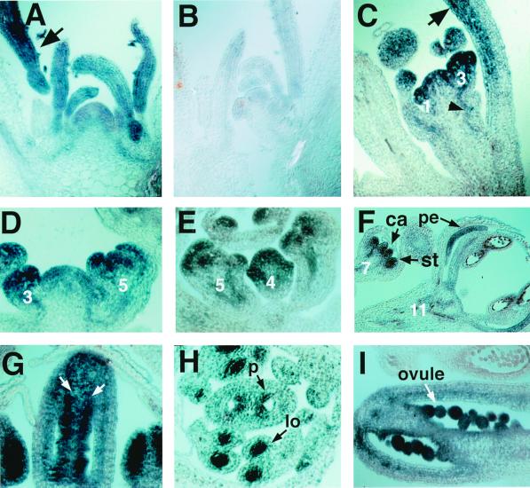
In situ expression pattern of LUG mRNA. (A) LUG expression in 14-day-old seedlings. A low level of LUG mRNA is present in the shoot apical meristem and in the first few young leaves. LUG mRNA level increases dramatically in more developed leaves (arrow). (B) LUG sense probe to 14-day-old seedlings. (C) LUG mRNA is detected in the secondary inflorescence meristem and in the stage 1 and 3 floral meristems (numbers indicate the stage of respective floral meristems; ref. 33). LUG mRNA is detected in vascular tissues (arrowhead). LUG mRNA level is also more abundant in the adaxial side of the cauline leaf (arrow). (D) LUG mRNA level is low in the inflorescence meristem but increases in young flowers (stages 3 and 5, respectively). (E) LUG is strongly expressed in the sepal primordia and the central dome of the stage 4 and 5 flowers. (F) At stage 7, LUG mRNA is not detected in sepals, but is present in the carpel (ca) and stamen (st) primordia. LUG mRNA is persistently detected in petals (pe) as shown in this stage 11 flower. (G) In stage 10 carpels, LUG mRNA is strongly expressed in the placenta/ovule primordia (arrows) and weakly expressed in the carpel valves. (H) A cross section of a stage 9 flower. A high level of LUG mRNA is detected in placenta (p) and locules (lo) of the anther. (I) LUG mRNA is detected in developing ovules at stage 12.

