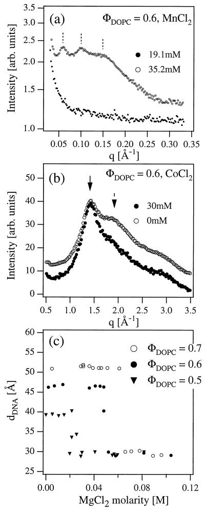
Figure 4.
(a) Synchrotron XRD measurements of the complex supernatant below (●) and above (○) the condensation transition in the presence of MnCl2. For M > M* the XRD pattern shows the existence of a multilamellar lipid phase of highly charged lipids expelled from the complexes during the 2D DNA condensation. (b) Wide-angle x-ray measurements of the complexes with CoCl2. Solid arrow indicates the peak caused by the fluid lipid chain packing and dashed arrow indicates the peak of water. (c) The onset of condensation occurs at lower values of MgCl2 concentration M* as the initial DNA interaxial spacing dDNA is decreased from 50.3 Å to 47.0 Å to 39.5 Å for ΦDOPC = 0.7, 0.6, and 0.5, respectively.

