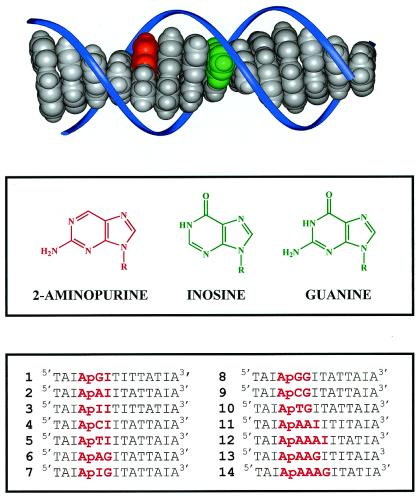
Figure 1.
(Top) Molecular model (INSIGHT II program) schematically illustrating the base pair stack within a DNA-duplex. The bases 2-aminopurine (Ap) and guanine (G) are depicted in red and green, respectively, and the sugar phosphate backbone is depicted schematically in blue. (Middle) Structures of 2-aminopurine, inosine, and guanine. (Bottom) The sequences of 14 DNA duplexes studied in our experiments. The complementary strands are not shown; Ap, as an analogue of adenine, is paired with thymine, and inosine is paired with cytosine.

