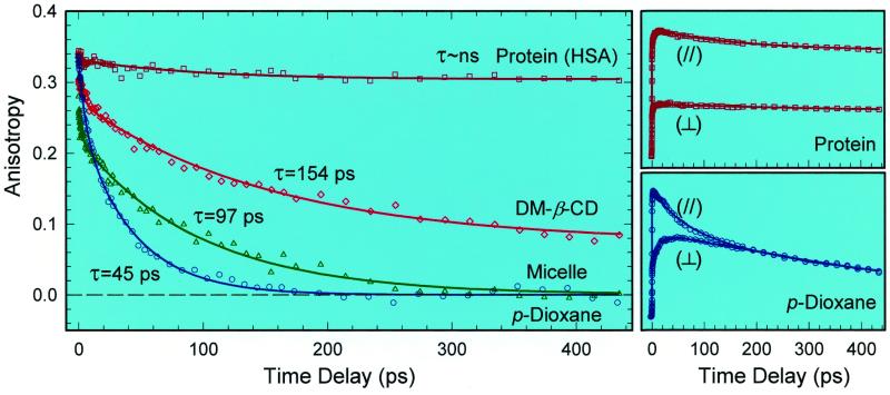
Figure 2.
Femtosecond-resolved fluorescence anisotropy evolution in four typical environments at 470-nm emission. Note the striking contrast between the free solvent and protein dynamics. The corresponding polarization-analyzed (I∥ and I⊥) transients for the HSA protein and p-dioxane are shown on the right.

