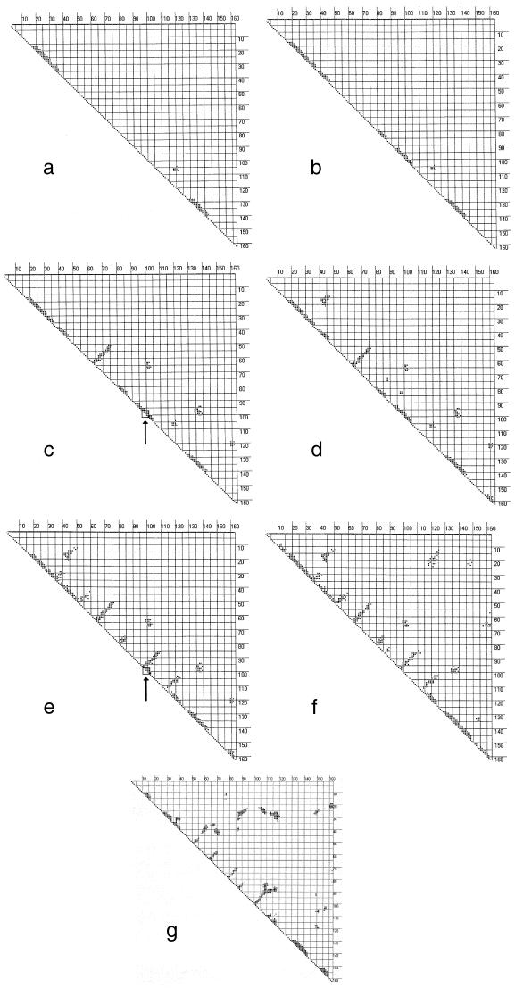
Figure 2.
Seven snapshots in the time evolution of the contact matrix for β-lactoglobulin obtained respectively (a–g) at 6.4 × 10⁻⁸, 3.0 × 10⁻⁷, 7.9 × 10⁻⁷, 1.8 × 10⁻⁵, 5.2 × 10⁻⁴, 1.0 × 10⁻³ s, and essentially the contact matrix for the native structure. The arrows in c and e indicate regions that show local topological invariance but structural multiplicity; that is, the relevant torsion angles remain in the same R-basins but change geometries.

