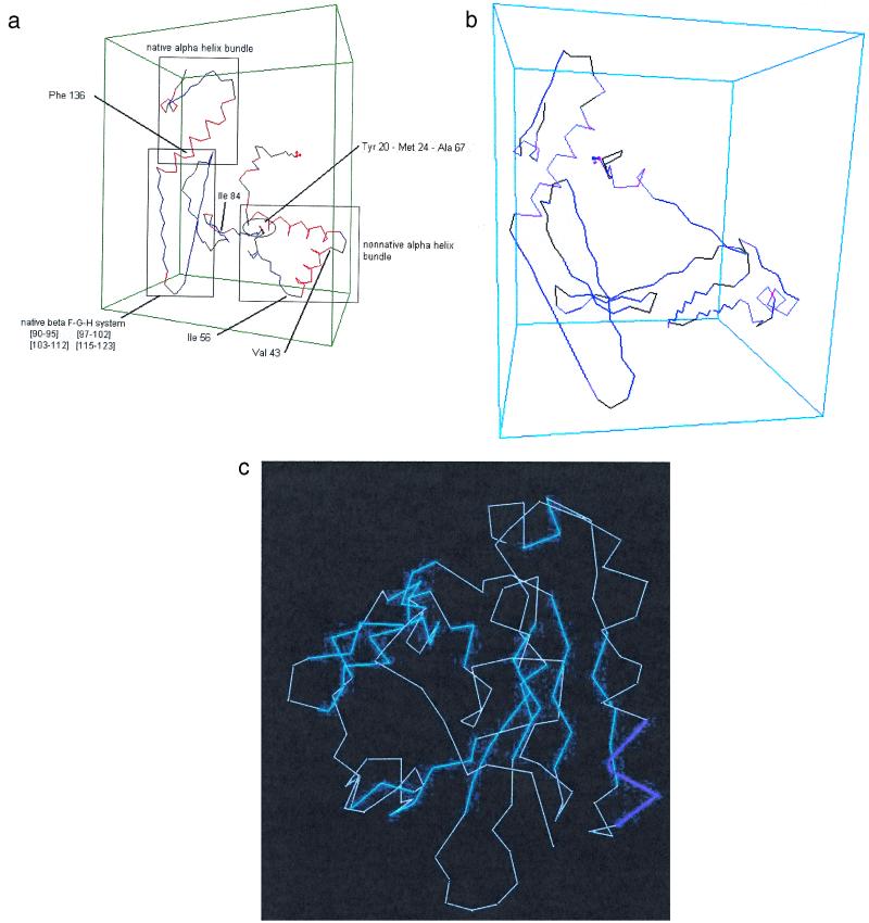
Figure 3.
Structures found for β-lactoglobulin at stages along the folding path of the contact maps of Fig. 2: (a) the structure corresponding to e; (b) the structure corresponding to f, not yet the native structure but after the transition from helical to β-structure in the 18–57 region. Sections shown in red are α-helices; sections in blue are β-structures. Sections in black are turns or random coils.

