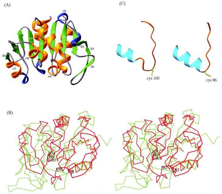
Figure 3.
Structural features of PH1704. (A) Ribbon diagram of PH1704. “H” and “S” refer to helix and strand, respectively. Green, gold, and blue code for β-strand, α-helix, and 310 helix, respectively. (B) The superposition of the Cα trace of amidotransferase domain of GMP synthetase (green) and that of PH1704 (red). The three members of the catalytic triad, Cys-86, His-181, and Glu-183 of the amidotransferase and Cys-100 and His-101 of PH1704 are labeled. (C) The “nucleophile elbows” in PH1704 (Left) and amidotransferase domain of GMP synthetase (Right).

