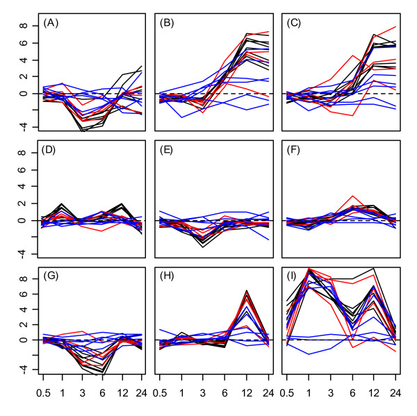
Figure 3.

Hsp20 expression response profiles in roots. Expression response profiles associated with Hsp20 proteins under (A) cold stress, (B) osmotic stress, (C) salt stress, (D) drought, (E) genotoxic stress, (F) oxidative stress, (G) ultraviolet-b light, (H) wounding, and (I) heat in root tissue. The cytoplasmic/nuclear Hsp20s (classes I – III) are represented by black lines. Plastidial, endoplasmic reticulum, and mitochondrial Hsp20s (classes P, ER, and M) are represented by red lines. Class I and Class P related Hsp20s are indicated by blue lines. The horizontal axis of each subplot corresponds to time points at which gene expression measurements were available under each stress treatment (0.5, 1, 3, 6, and 12 hrs.). The vertical axis of each subplot indicates log2 fold-change under a given stress treatment. A dotted horizontal line in each plot indicates a log2 fold-change of zero (no expression response to stress).
