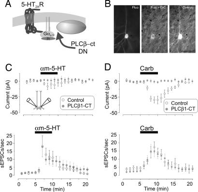
Fig. 2.
Single-cell inactivation of 5-HT2ARs by PLCβ-ct blocked the 5-HT2AR-induced inward current but not the increase in sEPSCs. (A) Diagram illustrating the site of action for the PLCβ-ct dominant negative. (B) Differential interference contrast (DIC)/fluorescence (Fluo) image of a neuron transfected with the PLCβ-ct dominant negative in an organotypic cortical slice (postnatal day 12; 3 days in vitro). (Scale bar: 50 μm.) (C) Paired recordings from neighboring neurons (Inset; n = 14) showing that the inward current elicited by αm-5-HT in control neurons was blocked in neurons expressing the PLCβ-ct construct (Upper; P < 0.05, paired Student's t test) but not the increase in sEPSCs (Lower; P = 0.59, paired Student's t test; n = 14 pairs). (D) Administration of carbachol (Carb) induced a slow inward current in control, nontransfected neurons (−29.9 ± 6.1 pA; n = 11) but not in neurons transfected with the PLCβ-ct construct (−0.4 ± 2.4 pA; n = 20; P < 0.01, unpaired Student's t test) (Upper). The ability of carbachol to induce an increase in sEPSCs was indistinguishable between control and transfected neurons (Lower; P = 0.41, unpaired Student's t test).

