Fig. 2.
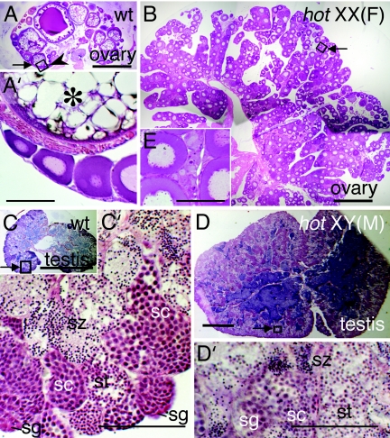
Histological sections of gonads from wild-type and hot-homozygous fish at 6 months after hatching. The specimens are sections of the tissues shown in Fig. 1K stained with hematoxylin-eosin. (A) Wild-type ovary. (B) XX hot-homozygous ovary. The arrowhead in A (enlarged in A′) indicates previtellogenic follicles, with which the immature follicles abundant in B share a similar appearance (enlarged in B′). An oocyte at the yolk formation stage is indicated by an asterisk in A′. (C) Wild-type testis. (C′) Enlargement of the boxed area in C (arrow), where spermatogonia (sg), spermatocytes (sc), spermatids (st), and spermatozoa (sz) are labeled according to the morphological criteria (39). (D′) Enlargement of the boxed area in D (arrow), showing sp, sc, st and sz. [Scale bars, 0.5 mm (A–D); 0.1 mm (A′–D′).]

