
Figure 1.
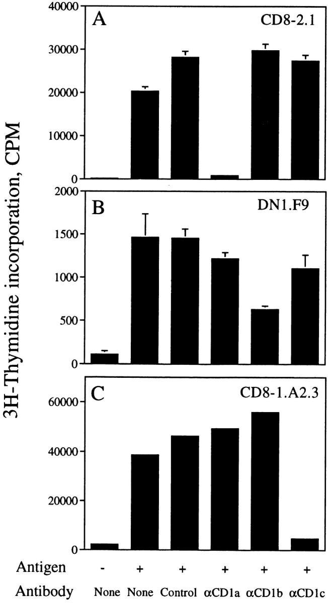
Specificity of T cell clones isolated from the CD8-2, DN1, and CD8-1 lines. CD8-2.1 (A), DN1.F9 (B), or CD8-1.A2.3 (C) T cells (5 × 10⁴/well) were cultured with CD1+ monocytes (5 × 10⁴/well) with or without antigen (silica fraction 90:10 at 2 μg/ml [A], mycolic acid at 40 μg/ml [B], or silica fraction 60:40 at 5 μg/ml [C]) for 72 h. The following mAbs were added at a final concentration of 20 μg/ml: P3 (IgG control), 10H3.9.3 (αCD1a), BCD1b3.1 (αCD1b), or F10/21A3.8 (αCD1c). [3H]Thymidine was added during the final 6 h of culture, after which the plates were harvested and [3H]thymidine incorporation measured in a liquid scintillation counter. Results are expressed as mean cpm ± SD of triplicate cultures (A and B). In C, results are from an experiment in which short-term clones were screened in singlicate. The pattern of antigen/CD1 recognition observed for these clones matches numerous experiments performed with the T cell lines.