
Figure 2.
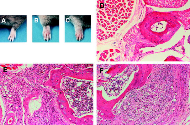
Clinical and histologic presentation of CIA in FcγRIIB−/− and DBA/1 mice. (A–C) The appearance of a normal forepaw from a CII-immunized wild-type mouse (A) contrasted with arthritic paws from an FcγRIIB−/− animal (B) and a positive control DBA/1 mouse (C). (D–F) Cross-sections of the forefoot from a normal wild-type mouse (D) compared with an arthritic joint from FcγRIIB−/− (E) and DBA/1 animals (F). Original magnifications: ×50 (D), ×80 (E), ×80 (F). D illustrates normal cartilage–bone without inflammation, whereas E and F show marked mononuclear cell infiltration with cartilage–bone destruction.