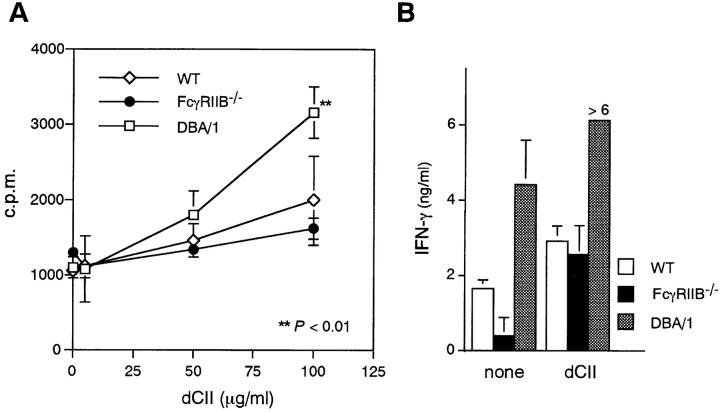
Figure 4.
Proliferation and IFN-γ production of anticollagen lymph node cells in response to CII. (A) Lymph node cells (5 × 10⁵/well) were stimulated in vitro with 5, 50, or 100 μg/ml heat-denatured CII (dCII) for 4 d. Proliferative response was determined by uptake of [3H]TdR pulsed for the final 18 h of culturing. (B) Each of the culture supernatants at the end of the experiment in A was collected and assessed for the IFN-γ content by ELISA. **P < 0.01 compared with wild-type mice.

