
Figure 9.
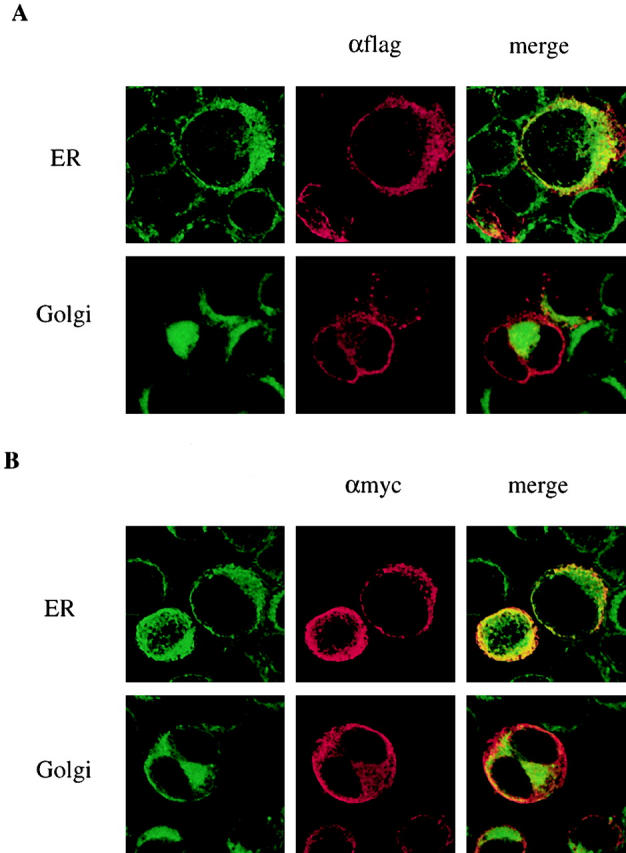
K1 localization. Expression vector containing (A) the flag-K1 or (B) the K1-myc was electroporated into BJAB cells. After 48 h, cells were fixed and reacted with mouse monoclonal anti-flag antibody (A) or anti-myc antibody (B). K1 protein was detected with anti–mouse secondary antibody conjugated with Alexa 568 (red). The ER and Golgi compartment were detected with Alexa 488–conjugated Con A (green) and BODIPY C5FL-ceramide (green), respectively. Localization of fluorescently labeled antibodies was visualized with a Leica TCS-SP laser scanning microscope. Yellow color indicates colocalization of the red and green labels. The data were reproduced in two independent experiments.