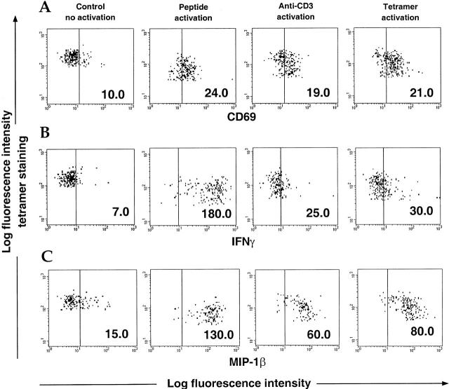
Figure 2.
Comparison among peptide, OKT3, and tetramer activation. PBMCs from a CMV-positive B7 donor were incubated for 12 h with brefeldin A in the presence of PBS (no activation control), specific CMV B7 peptide (10 μM), OKT3 (100 ng/ml), or CMV B7 tetramers. Tetramer staining was carried out after overnight incubation for the no activation control or before addition of the activators for the “activated” cells. Cells were stained for CD69 (A), IFN-γ (B), and MIP-1β (C) and analyzed by flow cytometry. Data show cells gated on the CMV B7 tetramer-positive population. Mean fluorescence intensity is shown for each condition.

