
Figure 3.
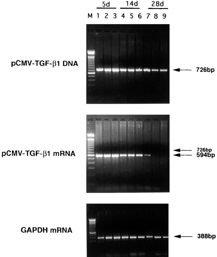
Analysis of pCMV-TGF-β1 DNA and mRNA in tissues by PCR and RT-PCR, respectively, at 5, 14, and 28 d after intranasal administration of pCMV-TGF-β1 and TNBS treatment. RT-PCR detects spliced mRNA message that differs in size from DNA and is thus not due to plasmid DNA contamination of endogenous TGF-β1 mRNA (described in text). Lane M, marker; lanes 1, 4, and 7, lung; lanes 2, 5, and 8, colon; lanes 3, 6, and 9, spleen; lanes 1–3, day 5; lanes 4–6, day 14; lanes 7–9, day 28. In TGF-β1 mRNA, a faint band of 726 bp was also detected because of the presence of unspliced message. One experiment representative of three independent experiments is shown.