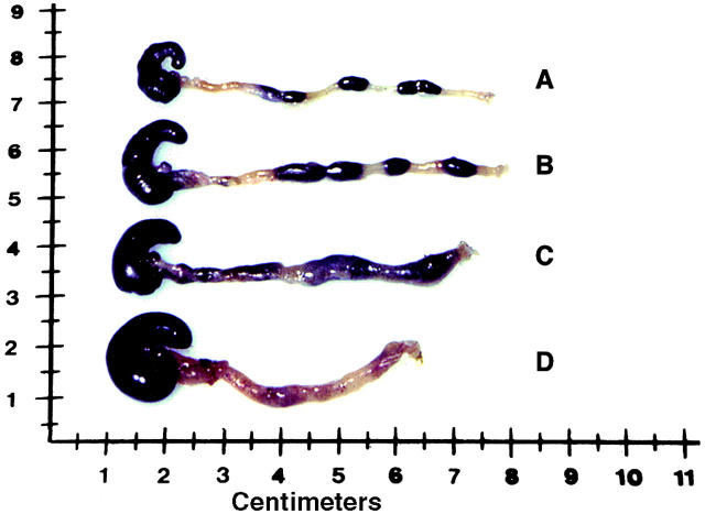
Figure 8.
Gross appearance of colons from SJL/J mice at 12 d after administration of TNBS on day 0, and after treatment with intranasal administration of pCMV-TGF-β1 and intraperitoneal administration of anti–IL-10 mAbs or rat control IgM on day 7. Representative colons from (A) a normal control mouse, (B) a mouse treated with pCMV-TGF-β1 (intranasally) and rat control IgM (intraperitoneally), (C) a mouse treated with pCMV-TGF-β1 (intranasally) and anti–IL-10 mAbs (intraperitoneally), and (D) nontreated established colitis.

