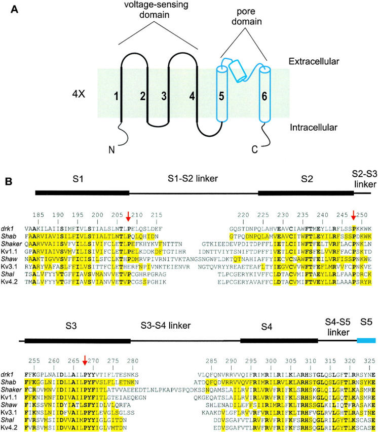
Figure 1.

Topology and sequence alignment of voltage-gated K+ channels. (A) Putative topology of a single subunit of the voltage-gated K+ channel with six putative transmembrane segments. Top is extracellular and the bottom is intracellular. In the tetramer, the coassembly of S5–S6 segments form the pore domain. The first four transmembrane segments form a single voltage-sensing domain with four of these domains surrounding the central pore domain. (B) Sequence alignment between the four classes of voltage-gated K+ channels in a region spanning four putative transmembrane segments (S1–S4) and linkers. Black bars above the sequence represent the approximate positions of the four transmembrane segments as indicated by the Kyte-Doolittle hydrophobicity analysis. Residue numbering is for the drk1 K+ channel. Yellow highlighting indicates similarity to drk1. Bold letters indicate residues that are highly conserved in all the voltage-gated K+ channels. Red arrows mark three highly conserved proline residues.
