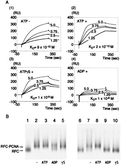
Figure 1.
Studies of the interaction between RFC and PCNA. (A) Surface plasmon resonance analyses. Indicated concentrations of RFC were injected into a sensor chip prefixed with about 1,000 resonance units of PCNA. All of the procedures were performed with 150 mM NaCl Running Buffer, with RFC as the analyte without nucleotides (1) or with 1 mM ATP (2), ATPγS (3), or ADP (4). KD values with SD obtained from three injections were (1 ± 0.4) × 10−9 M (2 ± 0.5) × 10−10 M (9 ± 2) × 10−10 M and (1 ± 0.8) × 10−8 M, respectively. (B) Gel mobility profiles of RFC and RFC-PCNA complexes without ATP (−, lanes 2 and 7), with ATP (lanes 3 and 8), with ADP (lanes 4 and 9), or with ATPγS (lanes 5 and 10) at 0.15 M (lanes 1–5) or 0.5 M (lanes 6–10) NaCl. Arrows indicate migration positions of silver-stained RFC and RFC-PCNA complex. In lanes 1 and 6, only RFC was applied. PCNA runs at the dye front and does not appear in this picture under these electrophoresis conditions.

