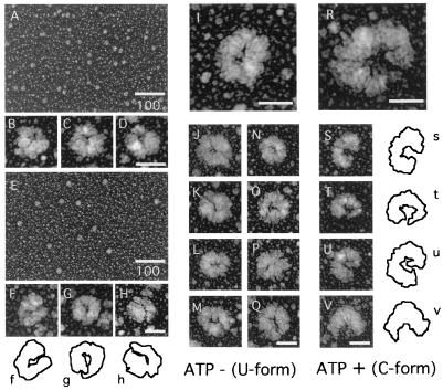
Figure 3.
Low-magnification images of PCNA (A) and RFC without ATP (E). B–D and F–H are high-power views from A and E. Standard length bars for A and E are 100 nm and for the others, 20 nm. The outlines of images for RFC in F–H are drawn below with f–h. Typical images of U- and C-form RFC obtained without and with ATP are shown in I and R, respectively. Examples of images of RFC in the U form without ATP (J–Q) and the C form with ATP (S–V) are shown. Outlines of images for the C form are drawn (Right) with s–v. The samples for I–V and A–H were prepared with and without crosslinking, respectively.

