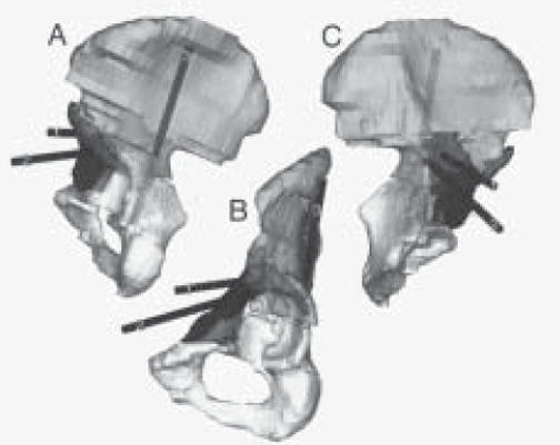
Figure 2.

Three rotated rendered 3-D CT images of the case study pelvis. Further segmentation allowed removal of the sacrum and contralateral hemi-pelvis. Three proposed screw trajectories (labeled 1, 2 and 3) have been added to the images and are visible through the translucent bone.
(A) Posterior view. (B) Transfemoral head inferior-lateral view. (C) Medial view. (1) Superior-posterior screw trajectory, (2) Superioranterior screw trajectory, (3) Posterior column screw trajectory.
