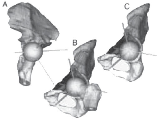
Figure 4.

Three rotated views of the segmented rendered 3-D pelvis with a sphere representing the proposed size and ideal location for an acetabular replacement component. (A) Same view as Figure 3A with proposed acetabular replacement trajectories instead of acetabular repair trajectories. (B) Same view as Figure 3B with sphere and femoral head. (C) Same view as Figure 3B with femoral head removed.
