Figure 5.
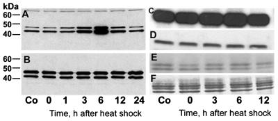
ERK1/2 shows increased activation after heat shock and hyperphosphorylates MBP but not τ. (A and B) Immunoblots of cerebral extracts from control (Co) and heat-shocked rats probed with the antibodies against activated and total ERK1/2, respectively. The phosphospecific antibody shows increased activation of ERK1/2 at 3 and 6 h after heat shock (A) but the total amount of ERK1/2 is unchanged (B). In A, note the equal loading of extract protein in each lane indicated by the nonspecific staining of a higher molecular weight protein. Audoradiographs (C and E) of Coomassie blue-stained (D and F) SDS/PAGE gels of kinase assay mixtures toward MBP (C and D) and τ (E and F) show hyperphosphorylation of MBP but not τ at 3 and 6 h after heat shock.

