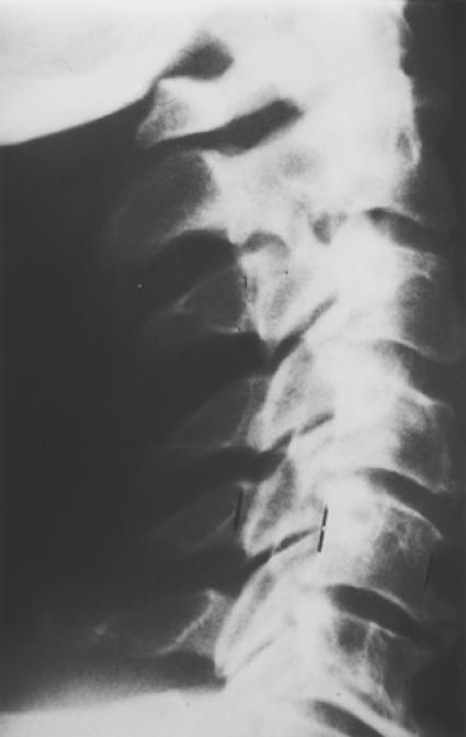
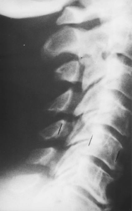
Figures 1-A and 1-B. Pre-and postoperative radiographs of a 65 year-old man with progressive myelopathy at the time of laminoplasty.
Fig. 1-A. Preoperative lateral radiograph demonstrating notable canal stenosis.

Fig. 1-B. Postoperative lateral radiograph showing a 7 millimeter expansion of the space-available-for-the-cord following laminoplasty using Hirabyashi's open-door technique.

