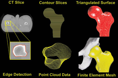
Figure 2.

Full sequence of pre-processing steps, beginning with edge detection of individual CT slices. Point cloud data, which record the spatial coordinates of individual points along the detected periosteal surface, result from the accumulation of contoured slices taken at 1mm increments. A triangulated surface was then fitted to the point cloud data for each side of the joint (femur here illustrated). Finally, an all-quadrilateral finite element mesh was projected onto the triangulated surface. The same sequence is used for both the acetabular and femoral sides.
