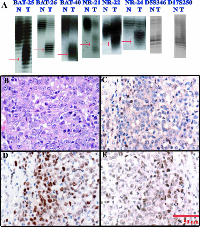
Figure 1.
Case 4 is an MSI+/hMLH1-immunonegative case with six of eight loci instability. A: Microsatellite instability in all six mononucleotide loci (red arrows, extra alleles in tumor genomic DNA) and normal-sized alleles in both normal and tumor using both dinucleotide markers D5S346 and D17S25. Note allelic shifts in DNA from tumor (T) samples compared with DNA from corresponding normal (N) tissue. B through E: Photomicrographs taken at 40× magnification. B: H&E showing mixed-type gastric cancer using the Carneiro system; C: hMLH1 immunostaining; D: hMSH2 immunostaining; E: hMSH6 immunostaining. Note the lack of tumor nuclear staining by hMLH1 and normal hMSH2 and hMSH6 immunostaining. Internal positive control immunostaining is demonstrated in nuclear staining of lymphocytes.

