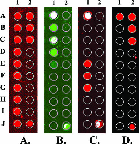
Figure 2.
Layout of the pathogen detection microarray probes and hybridization results. A: Image of one array hybridized with the Cy5-QC oligo. B: Image of one of the two replicate arrays in a microarray assay of blood spiked with B. anthracis (50 CFU/ml) labeled with Cy3. C: F. tularensis (50 CFU/ml) labeled with Cy5. D: Y. pseudotuberculosis (50 CFU/ml) labeled with Cy5.

