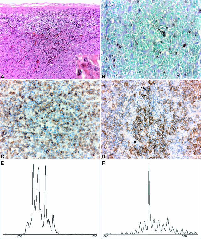
Figure 2.
Polymorphic B-cell PTLD-associated disease in case 27. A: Histology of LN. There was no recognizable lymph node architecture (HE; magnification, ×200). There were numerous blasts (inset) (HE; magnification, ×600) intermingled with an infiltrate of smaller lymphocytes. B: EBER-ISH showed scattered positively stained nuclei, indicating the presence of EBV RNA (magnification, ×400). C: CD3 staining of the predominantly small T cells. Occasionally, larger cells stained positively (magnification, ×400). D: CD20 stained the majority of the large blasts. Note the numerous mitotic figures (arrows) (magnification, ×400). E and F: PCR-based GeneScan analysis of LN DNA. Oligoclonal TCRB gene rearrangements could be identified (E), whereas a monoclonal IGH gene rearrangement was detected (F).

