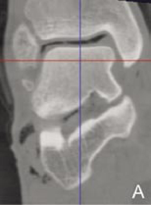
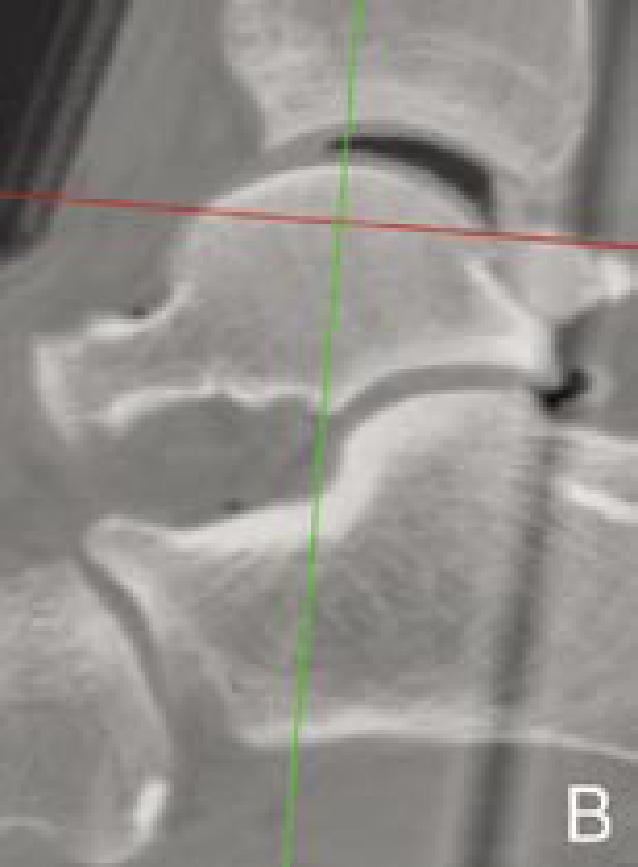
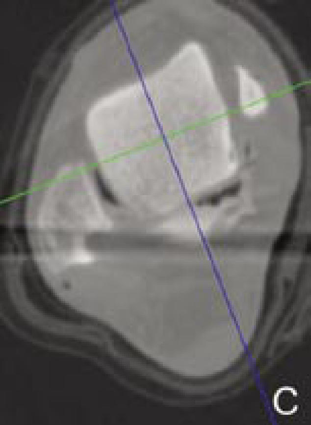
Figure 1.


Determination of local reference planes based on anatomical landmarks. The blue, green, and red axes on the images indicate the reference planes of the 3D-CT image coordinate system (sagittal, coronal, and transverse planes, respectively). First, in a coronal section passing the middle of the talar dome (A), the coordinate system is rotated and translated so that the red axis is parallel to the superior talar surface and the blue axis will bisect the talar dome width in the middle. Next, in the sagittal section (B), the coordinate system is rotated and translated so that the red axis is parallel to the line passing through the anterior and posterior ends of the articular surface, and the green line transects the talar dome at the middle. Finally, in the transverse section (C), the coordinate system is aligned so that the blue and green axes perpendicularly transect the talar dome.
