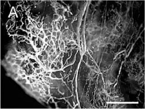
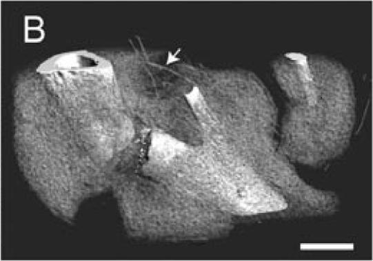
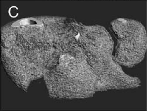
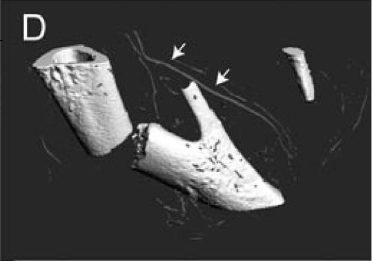
Figure 1.
Microfil blood vessel casting and micro-CT imaging. (A) Microfil casting demonstrated a network of blood vessels around the callus of a day 14 non-stabilized mouse tibia fracture. The same sample was scanned at 20 µm under micro-CT, and 3-dimensional images were reconstructed to show (B) the site of fracture, the mineralized callus, and blood vessels (arrow); (C) the mineralized callus only; and (D) the site of fracture and blood vessels (arrows). Scale bar = 1mm.


