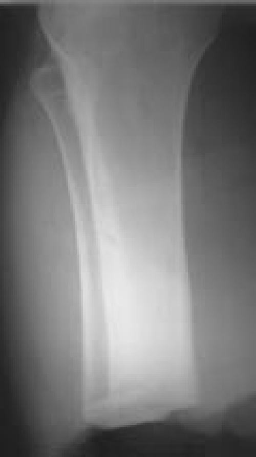
Figure 1A.

(Case 1) AP radiograph of a transtibial amputation with fibular cortical bone block.
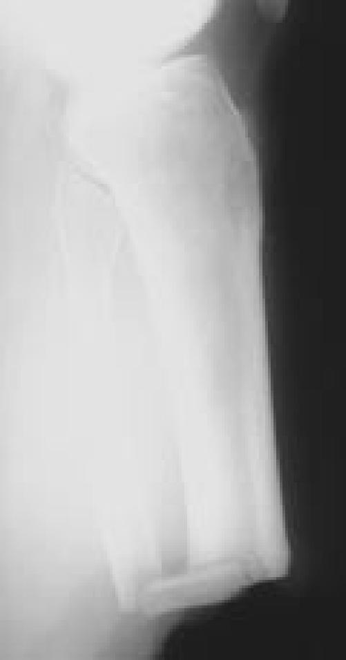
Figure 1B.

Lateral radiograph. Note the notching of the tibia to provide maximum bone surface for healing and the drill holes that were utilized for suture fixation.
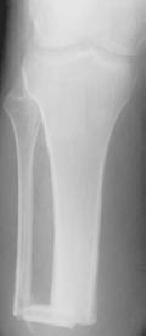
Figure 1C.

AP radiograph at 6 months demonstrates healing of the bone bridge.
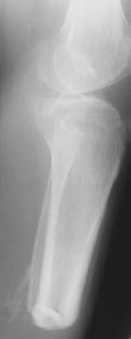
Figure 1D.

Lateral radiograph confirms healing. There is some ectopic bone formation posterior to the fibula.
