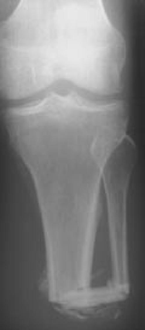
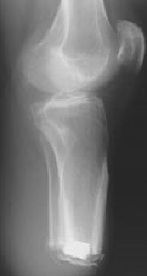
Figures 3A, B. (Case 5) AP and lateral radiographs of patient with a TTA with both fibular bone block and ostealperiosteal sleeve.
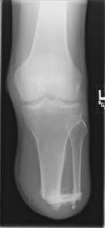
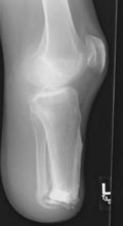
Figures 3C, D. AP and lateral radiographs at 6 months demonstrating good healing of the fibular bone block with additional new bone formation beneath the periosteal sleeve.
Figure 3A.

Figure 3B.


