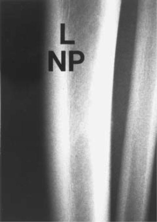
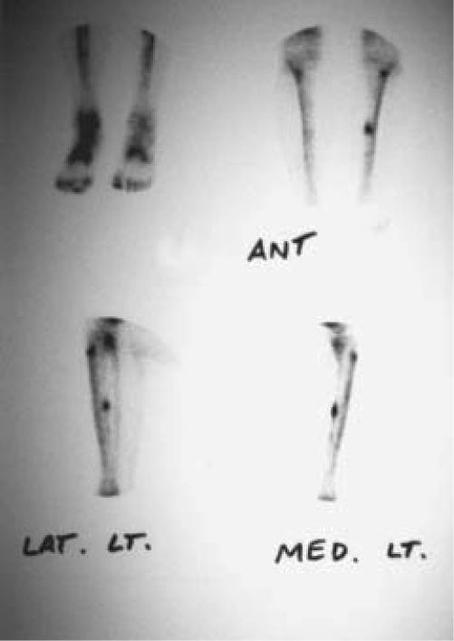
Figure 1.
N.P. is a 19 year old white male college level soccer play who complained of posterior-medial pain along the mid portion of the tibia.
Figure 1A.
(A) Plain radiographs of the tibia and fibula showing a small "cloud" of periosteal reaction consistent with a tibial stress fracture at the posterior-medial border of the left tibia. The accompanying bone scan confirmed the diagnosis.
Figure 1B.
(B) The bone scan is of both tibias. Increased uptake is noted along the posterior-medial border of the left tibia in the diaphysis.


