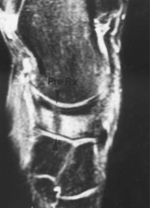
Figure 2.
W.B. is a 20 year old African American male, college level basketball player with an MRI documented tarsal navicular fracture. This image is from 12/16/97, before treatment was initiated. The stress fracture is visualized across the entire width of the tarsal navicular on the coronal view. The fracture line can be seen extending into subchondral bone.

