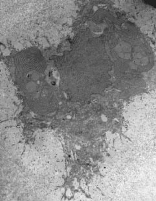
Figure 1.
Transmission electron micrograph of a pseudoachondroplastic dysplasia growth plate chondrocyte identifying alternately electron-lucent and electron-dense layers of material in the rough endoplasmic reticulum, the cytochemical hallmark pattern associated with pseudoachondroplastic dysplasia. Magnification = 3,900x.

