
Figure 7.
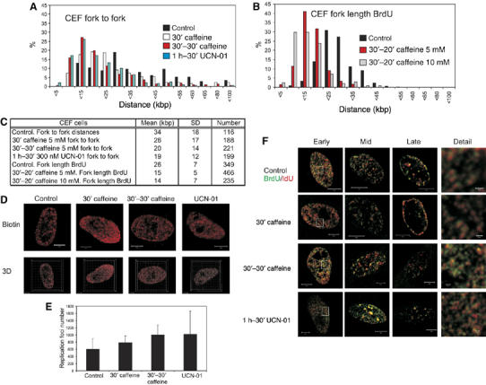
Chk1 regulates replication origin density in primary avian fibroblasts. Replication structures of chick embryo fibroblasts were visualised using the procedures described in Figure 2. Replicon size (A–C) was measured using the separation of sister forks (panel A) after labelling with biotin-dU and structures in untreated cells compared with those in cells treated with caffeine or UCN-01. BrdU labelling was also used to determine the rate of fork elongation (panel B) under the same conditions. Parameters defining average replicon structure under different conditions are shown (panel C). The number of active replication factories (D, E) and organisation of active replication foci (F) in cells treated with caffeine and UCN-01 were compared with untreated cells. Changes in the number of active DNA foci in treated cells were shown to be statistically significant using Mann–Whitney test: 30′ caffeine versus control (P<0.05; U-value 78); 30′–30′ caffeine versus control (P<0.005; U-value 88); UCN-01 versus control (P<0.02; U-value 82). Scale bars (5 and 0.5 μm in Detail) are shown on individual images.