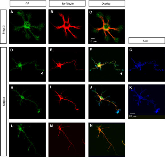
Figure 4.

Expression pattern of Gβ in various stages of cultured primary hippocampal neuron differentiation. Gβ distributes diffusely throughout the cell body and the minor processes in stage 2 neurons (A–C). As the neurons develop through stages 2–3 and reach stage 3 and adopt the well-differentiated neuronal phenotype, Gβ shows two distinct labeling patterns. Approximately 40% of stage 3 neurons (n>50) showed Gβ labeling in the central region of the axonal growth cone (D–K). An example of a stage 3 neuron that failed to show any detectable enhancement of Gβ in the growth cones is represented (L–N). Neurons were colabeled for Gβ (green in A, D, H and L), Tyr-Tubulin (red in B, E, I and M) and actin (blue in G and K). Overlayed images are shown in C, F, J and N. Scale bars equal 10 μm in panels (A–C) and 20 μm in panels (D–N).
